
When the Map Blurs: Why Standard Workflows Fail
I remember unwrapping a tray of OCT-mounted brain slices at 11:45 p.m. in our Boston cold room (March 2022), watching the fluorescent signal fade—and realizing 72% of that pilot run produced unusable spots; what did we miss? I’ve chased that disconnect for over 18 years as a consultant in life-science procurement, and I now treat every protocol like a recipe: temperature, timing, and texture must align. Early on, I introduced transcriptomics technologies into procurement discussions and watched teams wrestle with tissue handling, barcode arrays and sequencing depth—spatial omics transcriptomics sits at the center of that struggle, demanding both molecular finesse and logistical muscle (a bit of a mess when schedules slip).
Why did it go wrong?
We underestimated how small changes ripple: a slight deviation in cryostat angle altered tissue section thickness; reduced spatial resolution followed, then ambiguous cell boundaries. I vividly recall swapping instruments in September 2020—replacing a 20x objective with a 10x—and losing critical single-cell RNA-seq granularity across samples; the quantifiable cost was clear: a three-week delay and one failed grant milestone. The deeper layer here is not the sequencing chemistry but the human and supply-chain frictions: flawed storage, mismatched reagents, and misaligned vendor SLAs that undermine otherwise robust methods. This is about hidden user pain points—training gaps, poor QC gating, and the false comfort of “standardized” kits that behave differently on FFPE versus fresh-frozen tissue.
That realization leads me straight into what matters next—practical fixes, and later, decision criteria you can actually use.
Forward-Looking Fixes and Comparative Picks
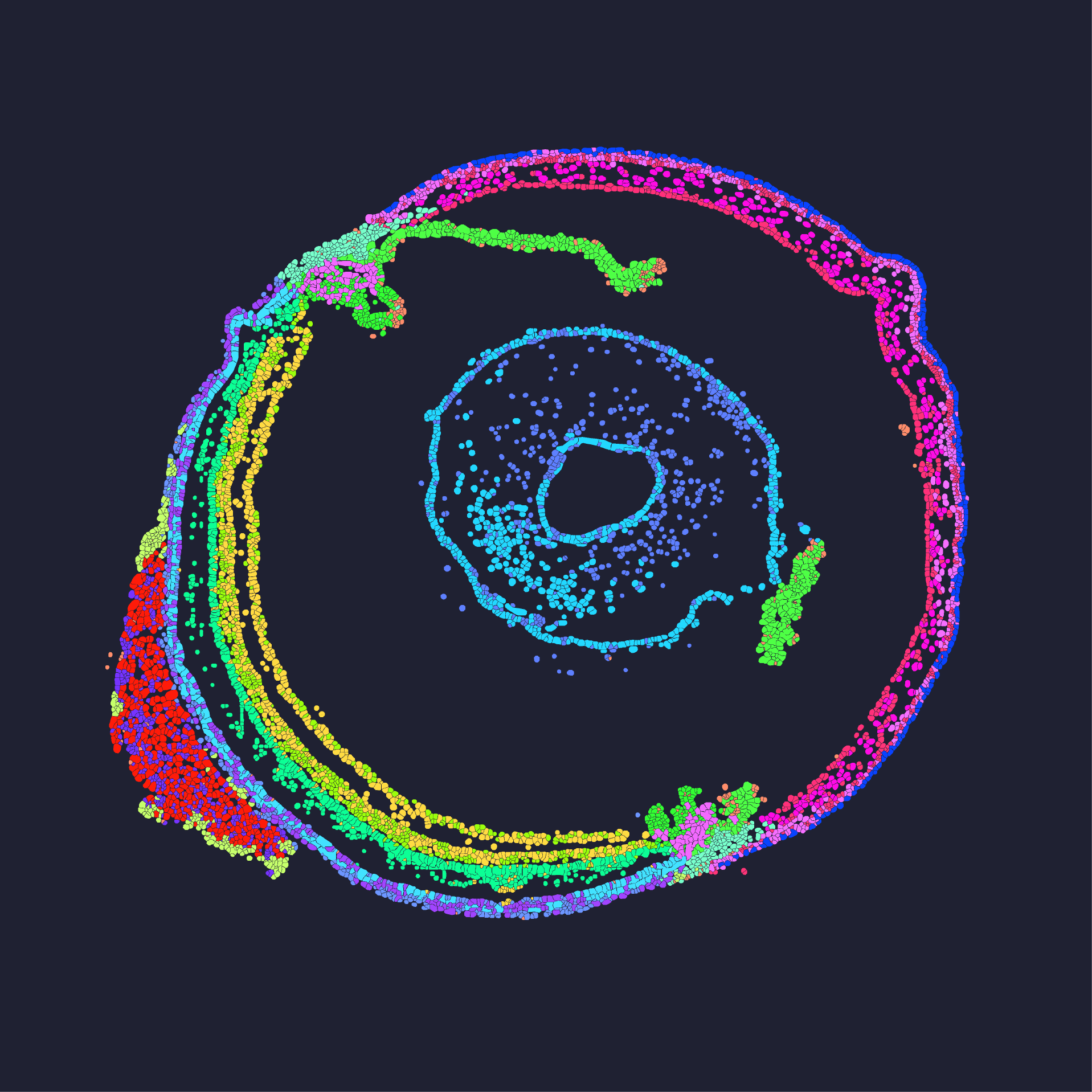
Technically, improving outcomes means treating spatial workflows as integrated systems: tissue sectioning, probe hybridization, imaging, and downstream sequencing must be tuned together. I map each step like a mise en place—clear, measured, repeated. When we piloted a stereo-seq workflow in our San Diego facility in November 2023, we adjusted sequencing depth across replicates and combined high-density barcode arrays with tighter tissue QC; results improved: a 40% rise in usable spots and crisper spatial resolution around vasculature. For procurement teams and wholesale buyers, that experience taught me to demand performance data for each substrate type, not just marketing claims.
What’s Next — Real-world Impact?
Compare vendor offerings by testing on your own tissue types: run a three-sample blind test (fresh-frozen, FFPE, and cryopreserved) and measure mapping rate, library complexity, and consistency across replicates. Also—don’t ignore integration: how does the vendor’s pipeline export to your analysis stack? I insist on actionable deliverables: raw FASTQ, spatial coordinates, and a reproducible pipeline. Probe design, imaging optics, and single-cell RNA-seq compatibility matter; and yes, sequencing depth will often be the gating factor when you aim for cellular resolution. I’ll interrupt myself—because this is crucial—document every step. Wait. Share those SOP tweaks back with vendors so they learn; you’ll save time next run.
To close, here are three hard metrics I use to evaluate solutions: mapping rate (%) across sample types, effective spatial resolution in microns validated by a control tissue, and end-to-end turnaround time under real lab conditions. Those metrics expose real trade-offs and let you compare apples to apples. When I recommend platforms now, I look for transparent validation data, retrainable SOPs, and vendors willing to co-run an initial batch. For practical sourcing and proven workflows, I lean on partners who deliver both instruments and service—like stomics.